
A laceration of the nail matrix that is poorly assessed in the first hours determines the final morphology of the nail plate. The fibrous scar that forms in the absence of precise anatomical repair becomes the permanent mold of a ridged, split, or deviated nail. Here, we discuss the mechanisms that render certain deformities irreversible, the available corrective actions, and concrete measures for secondary prevention.
Matrix fibrous scar: the mechanism of permanent dystrophies
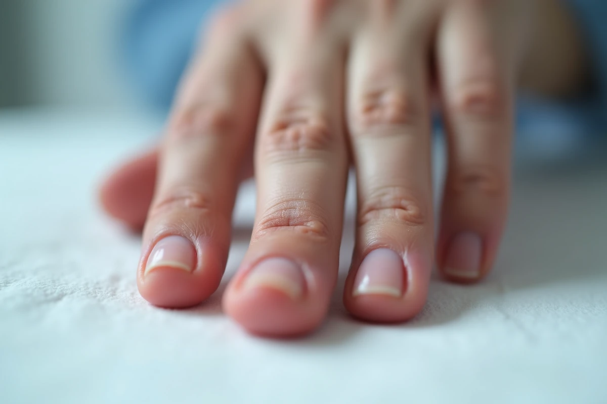
The nail matrix functions as a specialized germinative epithelium. When trauma destroys a portion of this tissue, the injured area is replaced by collagenous scar tissue devoid of keratinogenic capacity. This fibrous tissue does not produce a nail plate, creating a longitudinal split or localized thinning with each regrowth cycle.
See also : How to Prevent Grass from Sticking Under the Mower: Simple and Effective Tips
The distinction between proximal matrix and distal matrix is crucial. Damage to the proximal matrix alters the dorsal surface of the nail plate, producing striations or palpable ridges. A lesion of the distal matrix, on the other hand, modifies the ventral side, resulting in a nail that lifts or thickens irregularly.
What freezes the deformation is the delay. Early reconstruction after matrix laceration allows for the repositioning of the germinative edges before fibrosis organizes. Beyond this stage, the fibrous scar traps the remaining stem cells in a disordered architecture. We regularly observe in consultations dystrophies that could have been avoided if the initial repair had included a magnified examination and precise anatomical confrontation of the matrix fragments.
You may also like : How to Effectively Install Your Induction Cooktop: Tips and Methods
A trauma to the nail matrix that is managed late or superficially thus leaves a permanent functional defect in the germinative tissue, and it is this defect, not the initial trauma, that produces the visible deformation months later.
Matrix repair and catch-up graft: correcting an established nail dystrophy
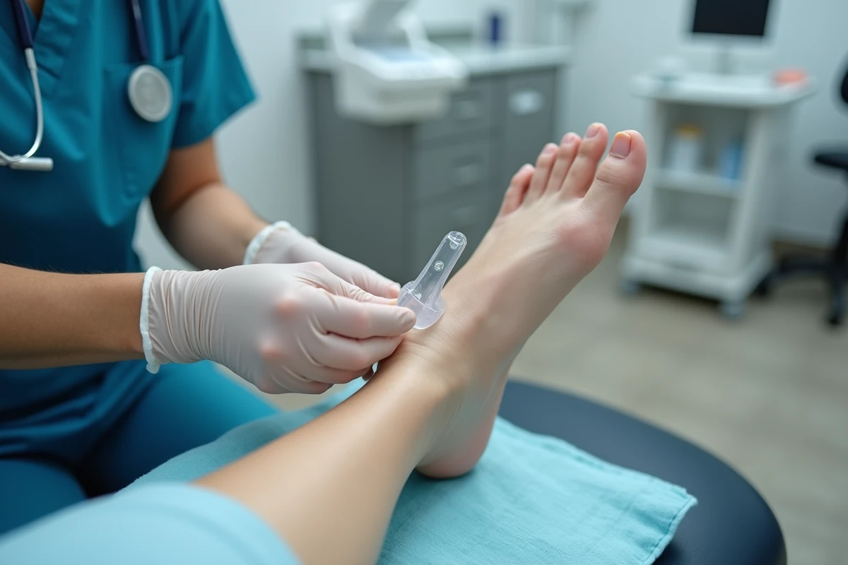
When the dystrophy is already established, two surgical options stand out depending on the extent of the matrix damage.
Scar revision under magnification
For isolated longitudinal splits caused by a linear fibrous scar, the revision involves excising the scar tract and bringing the healthy edges of the matrix closer together. The procedure is performed under binocular magnification or operating microscope.
The suture must be performed with a fine absorbable thread (type 6-0 or 7-0) to avoid any tension on the germinative tissue. A suture that is too tight or a thread that is too thick creates a new area of local ischemia, and thus a new scar.
Graft of healthy matrix tissue
When the matrix destruction is too extensive for a simple revision, a graft of matrix taken from a donor toe constitutes a catch-up option described in recent publications. The graft provides germinative cells capable of resuming orderly keratinization. The donor site is usually a toe from which partial matrix loss remains functionally acceptable.
This technique does not guarantee a nail with a normal appearance. It aims to restore a continuous nail plate, without splits or painful relief, which already represents a significant functional and aesthetic gain compared to an untreated dystrophy.
Secondary prevention after trauma: protecting the matrix during healing
The prevention of deformities does not stop at the emergency room. The matrix healing phase, which extends over several weeks, remains vulnerable to repeated microtraumas.
- Wearing a protective splint during the first weeks limits mechanical shocks to the matrix under repair, especially in patients exposed to manual activity or wearing rigid shoes
- Repositioning the eponychium after suturing prevents the formation of adhesions between the roof of the proximal nail fold and the nail bed, adhesions that deflect the regrowth
- The original nail plate, even if fractured, serves as a natural biological splint when replaced under the eponychium: it keeps the dead space open and guides the new growth in the correct axis
- Monitoring for superinfections is essential, as an infection of the nail bed during the healing phase worsens fibrosis and extends the area of matrix destruction
Warning signs and decision for partial matricectomy
Not all dystrophic nails warrant an attempt at reconstruction. We recommend a delayed evaluation of the trauma (at least two complete regrowth cycles) before indicating surgical intervention, as some minor deformities may spontaneously diminish as the healed matrix remodels.
On the other hand, several situations point towards intervention:
- A persistent longitudinal split that catches on fabrics or causes pain on contact
- A localized thickening (post-traumatic onychogryphosis) making shoe wearing painful
- An anarchic regrowth with recurrent ingrowth of the lateral edge
When the residual matrix produces only a painful and non-functional nail plate, partial or total matricectomy becomes a legitimate option. It permanently eliminates regrowth in the affected area, thus removing the pain-deformation-superinfection cycle. This decision is made on a case-by-case basis, weighing the aesthetic loss against the functional benefit.
The timing of the initial repair remains the main prognostic factor. A matrix reconstructed in the first hours with precise anatomical confrontation under magnification offers the best chances for orderly regrowth. Conversely, a quick suture with the naked eye, without exploration of the proximal nail fold, exposes to a dystrophy that only a catch-up graft can partially correct months later.